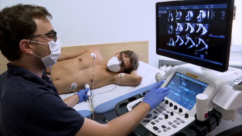
A promising new research study directed by Children’s Hospital Colorado in Aurora, Colo., viewed that three-dimensional (3D) echocardiography brought about more precise pacemaker lead placement when contrasted with X-ray technology. The study was presented at the 34th Annual Scientific Sessions of the American Society of Echocardiography (ASE), which took place in National Harbor, Maryland, from June 23 to 26.
Advances in echocardiography have led to more precise pacemaker placement, according to research presented at the 34th Annual Scientific Sessions of the American Society of Echocardiography (ASE 2023), which took place in National Harbor, Maryland, from June 23 to June 26, 2023.
The study, named 3D Echocardiography Guidance for Pacemaker Lead Placement Improves Accuracy of Lead Placement and Reduces QRS Duration Compared to Fluoroscopic Guidance, was introduced June 25 during ASE 2023, and one of almost 400 abstract poster presentations included during the event to feature proceeding with innovations in the field of cardiovascular ultrasound.
Leads, or flexible, insulated wires, are used in some pacemakers to deliver electrical pulses that help hearts beat normally. As summed up in an online ASE 2023 news summary, the current standard practice for setting pacemaker leads is fluoroscopic/X-ray guidance. Notwithstanding, a new research study led by Children’s Hospital Colorado in Aurora, Colo., saw that as three-dimensional (3D) echocardiography brought about more exact pacemaker lead placement when contrasted with X-ray technology.
Lead author Dale Burkett, MD, Assistant Professor in Pediatrics-Cardiology at Children’s Hospital Colorado, explained that pacemaker leads can better mimic the heart’s normal electrical conduction and lower the risk of heart dysfunction in the long run when placed in the right location. Additionally, according to the research spotlight section of the ASE, 3D echocardiography reduces radiation exposure during the procedure for both the patient and the cardiology staff.
“By using 3D echocardiography guidance, we are able to better visualize pacemaker leads as they move through the heart and guide them to where they are intended to go,” said Burkett, adding, “Our work demonstrates that with advances in echocardiography, we can provide higher quality care for our patients who need permanent pacemakers for heart rhythm management.”
The following is a summary of the presented methodology, results, and conclusion:
The team described the procedure in detail, noting that 3DE was used to direct pacemaker leads to the desired locations, where they were secured, in 59 patients; Only when necessary for lead movement, lead securing, or slack adjustment was fluoroscopy used sparingly. Echocardiography and CT or MRI were used to locate the lead in those with it. RV leads had their paced QRS duration measured. Wilcoxon rank sum, Pearson’s chi-square, or Fisher’s exact tests were used to compare the cases to 72 previous control pacemaker cases that only used fluoroscopy for the lead implant.
Recorded as a hard copy of their outcomes, the creators offered this: Using 3DE guidance, echocardiography determined that 78.4 percent of RV leads were located in the septum, 17.7 percent in the junction between the septum and free wall, and only 3.9 percent were located in the free wall. By contrast, 29.8 percent, 12.8%, and 55.4 percent were located in the free wall using fluoroscopic guidance.
They added that this pattern was likewise found in the set number of patients that had a resulting CT or X-ray. Of the two RV leads put in the free-wall while utilizing 3DE direction, one was an ICD-just lead, and in the other, the ICD+pacemaker lead was directed to the septum however pacing boundaries were not decent and consequently the free-wall was utilized. Patients who received 3DE guidance with the lead implanted in the septum had a significantly shorter QRS duration (129 milliseconds, IQR 117-143) than those who received fluoroscopic guidance alone (141.5 milliseconds, IQR 134-148) (P=0.031).
The researchers came to the conclusion that, in comparison to fluoroscopic guidance, 3DE guidance for pacemaker lead placement is more accurate in placing the lead in the septum and has a shorter QRS duration. They also stated that 3DE guidance for lead placement has the potential to lower the long-term risk of pacemaker-induced cardiomyopathy and ought to be taken into consideration for all pacemaker lead placements.
A finalist in the 2023 Arthur E. Weyman Young Investigator’s Award Competition, the study was presented. The following researchers collaborated with Burkett on the study: Kathryn Collins, MD, Dustin Nash, MD, Martin Runciman, MBBS, Johannes von Alvensleben, MD, all of Children’s Hospital Colorado, (Aurora, CO); and Pei-Ni Jone, MD, of Lurie Children’s Hospital of Chicago (Chicago, IL).


 Cardiology2 weeks ago
 Cardiology2 weeks ago
 Neurology2 weeks ago
 Diabetology2 weeks ago
 Cardiology1 week ago
 Neurology2 weeks ago
 Neurology1 week ago