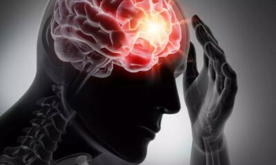
Is there ever a moment when you know exactly what you wanted to tell a buddy, but moments later you forget? According to experts, this can indicate a poor working memory, which is the capacity to retain information while completing other tasks for brief intervals of time.
Patients with dementia and Alzheimer’s disease frequently exhibit a working memory deficit, as does moderate cognitive impairment (MCI). Scientists are currently working to figure out the mechanics underlying this.
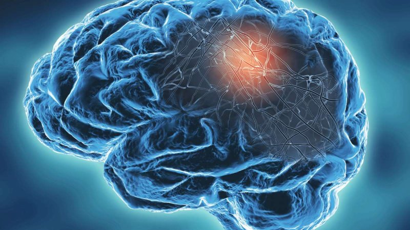
Researchers have found a mechanism that, even while we sleep, reduces metabolic expenses and builds memories. According to a UCLA statement, this process functions in the entorhinal cortex, which is the starting point of Alzheimer’s disease and a critical area of the brain for learning and memory.
Understanding the brain’s working memory
Researchers at UCLA Health sought to comprehend the metabolic cost of working memory tasks in the neocortex, the outermost layer of the brain. They paid special attention to how the entorhinal cortex and neocortex interact when under sleep anesthesia. The entorhinal cortex is where Alzheimer’s disease initially manifests.
This knowledge may shed light on memory issues like dementia, Alzheimer’s disease, and moderate cognitive impairment.
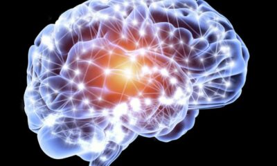
“It’s therefore critical to understand what kind of magic happens in the cortico-entorhinal network when the neocortex speaks to the entorhinal cortex which turns it into working memory,” stated corresponding author Mayank Mehta, a neurophysicist and head of the W. M. Keck Center for Neurophysics and the Center for Physics of Life at UCLA.
“It could provide an early diagnostic of Alzheimer’s disease and related dementia and mild cognitive impairment.”
Therefore, researchers developed a novel method known as the “mathematical microscope” to enable an early identification of such a memory or cognitive deficit.
The brain’s trillions of connections and billions of neurons were interpreted using a quantitative method.
“To tackle this seemingly impossible challenge of devising a simple theory that can still explain the experimental data of memory dynamics in vivo data with high precision, we hypothesized that cortico-entorhinal dialog, and memory magic, will occur even when the subjects are sleeping, or anesthetized,” stated Dr. Krishna Choudhary, the lead author of the study.
“Just like a car behaves when it’s idling or going at 70 mph,” added Dr. Choudhary.
Complex interactions are simplified by quantitative solutions.
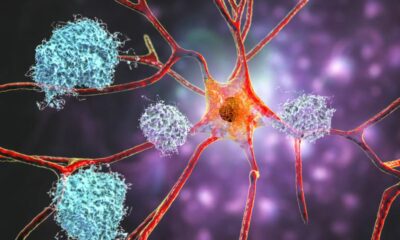
The intricate relationships between billions of neurons in the brain while it is asleep or under anesthesia have also been simplified by researchers. They believed that the dynamics of the entire system could only be represented by two neurons.
These two neurons were selected to symbolize the intensity of recurrent connections inside the entorhinal cortex as well as the strength of input coming from the neocortex.
Mehta stated, “These are just fascinating mathematical games, not a solid understanding of memory-making magic, if we test our theory quantitatively on data in vivo.”
According to Hahn, the entorhinal cortex is a complex circuit.“To really test the theory we needed experimental techniques that can not only measure the neural activity with high precision, but also determine the precise anatomical identity of the neuron.”
As a result, the “mathematical microscope” that was developed sought to grasp the complex processes involved in memory creation in the brain, improving our knowledge of how memories are formed while using less metabolic resources.
Additionally, the model unveiled a brand-new category of memory state known as “spontaneous persistent inactivity,” which boosts memory capacity with little energy usage.


 Diabetology2 weeks ago
 Diabetology2 weeks ago