General Medicine
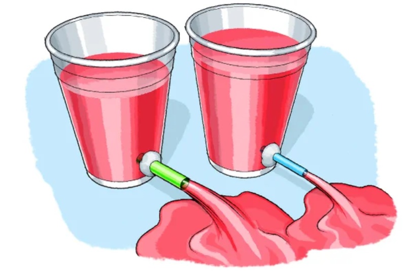
Breaking Down Artery Model Construction: Researchers Analyze Materials Used in Testing

Diabetology6 hours ago
Ayurvedic Remedies to Manage Type 2 Diabetes Effectively

Diabetology7 hours ago
Scientists Discover Sweet and Simple Way to Reduce Diabetes Risk — No Medicine Required

Diabetology4 days ago
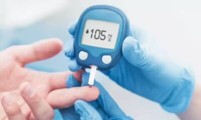
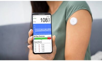
How AI Is Revolutionizing Diabetes Care: From Glucose Monitoring to Prevention

Pulmonology2 years ago
Lung growth invading Treg have different transcriptional profiles and capability connected to designated spot bar reaction

Dermatology2 years ago
Psoriasis be treatment by adjuvant Salicylic acid, ceramides as this containing lotions

General Medicine2 years ago
The CDC cautions physicians to be vigilant about uncommon, fatal flesh-eating germs
-
General Medicine4 days ago
Advancing Medical Innovations: Exploring the Breakthroughs of Microsurgery
-
 Diabetology1 week ago
Your Natural Solution for Blood Sugar Control
-
 Diabetology4 days ago
How AI Is Revolutionizing Diabetes Care: From Glucose Monitoring to Prevention
-
 Diabetology6 hours ago
Ayurvedic Remedies to Manage Type 2 Diabetes Effectively
-
 Diabetology7 hours ago
Scientists Discover Sweet and Simple Way to Reduce Diabetes Risk — No Medicine Required